
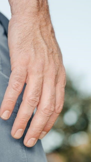
Surface Anatomy of the Hand
The hand’s surface anatomy includes the thumb, opposing four fingers, palmar aponeurosis, and fascia․ The thenar eminence (thumb) and hypothenar (little finger) are key functional regions․
1․1․ Bones and Joints
The hand contains 27 bones, with 8 carpal bones in the wrist, 5 metacarpal bones forming the palm, and 14 phalanges in the fingers and thumb․ Each finger has 3 phalanges, while the thumb has 2, allowing precise movement․ The metacarpophalangeal joints (MCP) are knuckle joints connecting metacarpals to phalanges, enabling flexion and extension․ The interphalangeal joints (IP) link phalanges, facilitating finger bending․ The thumb’s carpometacarpal joint (CMC) permits opposition, a unique human feature․ These bones and joints create a flexible, adaptive system for gripping and manipulating objects․
1․2․ Muscles of the Hand
The hand’s musculature includes intrinsic and extrinsic muscles․ Intrinsic muscles, such as the thenar (thumb), hypothenar (little finger), and interossei, are located within the hand, controlling fine movements․ They attach to carpal bones and phalanges, enabling actions like thumb opposition and finger adduction․ Extrinsic muscles in the forearm, including flexors and extensors, regulate wrist and finger movement via long tendons․ The flexor pollicis longus is vital for thumb flexion, while lumbricals and palmar interossei facilitate precise grip and coordination․
Anatomy of the Fingers
Fingers consist of three phalanges (proximal, intermediate, distal) and two interphalangeal joints, enabling flexion and extension․ The thumb has two phalanges and one joint, facilitating opposition․ Flexor and extensor tendons regulate movement․
2․1․ Phalanges and Their Structure
The phalanges are the bones forming the fingers and thumb․ Each finger contains three phalanges (proximal, intermediate, distal), while the thumb has two․ These bones are connected by joints, enabling flexion and extension․ The proximal phalanx is the base, attaching to the metacarpals, while the distal phalanx forms the fingertip․ The structure of phalanges varies slightly between fingers and thumb, with the thumb’s phalanges adapted for opposition․ This arrangement supports the hand’s dexterity and grip capabilities, essential for precise movements․
2․2․ Flexor and Extensor Tendons
Flexor tendons enable finger flexion, passing through the carpal tunnel․ The flexor digitorum superficialis and profundus tendons control finger movement, while the flexor pollicis longus governs thumb flexion․ Extensor tendons, located on the hand’s dorsal side, facilitate finger and thumb extension․ These tendons work in tandem with muscles in the forearm, enabling precise grip and pinch movements․ Their coordinated function is vital for hand dexterity and fine motor activities, such as grasping objects or performing intricate tasks․
Thumb Anatomy
The thumb comprises two phalanges and a carpometacarpal joint, allowing opposition and flexibility․ Its thenar muscles enable precise movements, while intrinsic muscles enhance dexterity and grip functionality․
3․1․ Opposing Thumb Mechanism
The opposing thumb mechanism is a unique anatomical feature enabling the thumb to touch all other fingers․ This function is facilitated by the carpometacarpal joint (CMC), which allows thumb opposition․ The CMC joint is a saddle-type joint, providing a wide range of motion, including flexion, extension, abduction, and adduction․ The thenar muscles, particularly the opponens pollicis, play a crucial role in this mechanism․ The flexor pollicis longus and extensor pollicis brevis muscles further enhance thumb mobility, making precise grip and pinch movements possible․ This mechanism is essential for grasping and manipulating objects, distinguishing human hand functionality․
3․2․ Thenar Muscles and Their Function
The thenar muscles, located at the thumb’s base, include the abductor pollicis brevis, flexor pollicis brevis, and opponens pollicis․ These muscles originate from the scaphoid, trapezium, and flexor retinaculum․ The abductor pollicis brevis facilitates thumb abduction, while the flexor pollicis brevis enables flexion at the metacarpophalangeal joint․ The opponens pollicis is crucial for thumb opposition, allowing the thumb to touch all fingers․ Together, these muscles enhance grip, pinch, and precise thumb movements, essential for hand functionality and dexterity․

Joint Structure and Function
The hand’s joints include metacarpophalangeal (MCP) and carpometacarpal (CMC) joints, enabling flexion, extension, and opposition movements․ The thumb’s CMC joint allows circumduction, while MCP joints facilitate finger dexterity․
4․1․ Metacarpophalangeal Joints (MCP)
The metacarpophalangeal joints (MCP) are located between the metacarpal bones and the proximal phalanges of the fingers․ These joints are crucial for finger movements, including flexion, extension, abduction, and adduction․ The MCP joints are stabilized by a joint capsule, collateral ligaments, and a volar plate․ They are classified as condyloid joints, allowing a wide range of motion․ Each MCP joint in the fingers has a similar structure, while the thumb’s MCP joint is more mobile due to its unique role in opposition and grip dynamics․
4․2․ Carpometacarpal Joint (CMC) of the Thumb
The carpometacarpal joint (CMC) of the thumb is a saddle-shaped joint that connects the first metacarpal bone to the trapezium․ It enables opposition, circumduction, and limited flexion-extension․ The joint is stabilized by ligaments and a fibrous capsule․ Its unique anatomy allows the thumb to oppose the fingers, facilitating grip and pinch․ The CMC joint is crucial for fine motor skills and hand dexterity, making it a key structure in thumb functionality and overall hand mechanics․
Blood Supply to the Hand
The hand receives blood from the radial and ulnar arteries, forming the superficial and deep palmar arches․ These arches supply the palmar surface, fingers, and thumb․ The fingers receive blood from palmar digital arteries, while the thumb is supplied by the princeps pollicis artery․ A collateral circulation ensures adequate blood flow, even when one major artery is compromised․
5․1․ Arteries and Veins in the Fingers
The fingers receive their blood supply primarily from the palmar digital arteries, which originate from the superficial palmar arch․ These arteries run along the palmar surface of the fingers, providing oxygenated blood to the skin, muscles, and tendons․ The dorsal digital arteries supply the extensor tendons and nail beds․ Venous blood returns through palmar and dorsal venous networks, draining into the palmar and dorsal venous arches․ This dual circulation ensures consistent blood flow and supports the fingers’ high mobility and sensitivity․
5․2․ Vascular Supply to the Thumb
The thumb’s vascular supply is primarily provided by the princeps pollicis artery, a branch of the radial artery․ This artery runs along the palmar surface of the thumb, supplying the skin, muscles, and tendons․ The proper palmar digital artery also contributes, ensuring rich blood flow․ Venous drainage occurs through the palmar and dorsal venous networks, which converge into the venous arches of the hand․ This dual circulation ensures the thumb’s high functionality and sensitivity, supporting its complex movements and grasp capabilities․

Nerve Innervation
The hand’s nerve innervation is provided by the median, ulnar, and radial nerves․ The median nerve supplies the thumb, index, and middle fingers, while the ulnar nerve supplies the ring and little fingers․ The radial nerve contributes to the dorsal wrist and finger areas․ Sensory and motor functions are distributed to enable precise movements and sensitivity․
6․1․ Sensory and Motor Nerves in the Fingers
The fingers receive sensory and motor innervation primarily from the median and ulnar nerves․ The median nerve supplies the thumb, index, and middle fingers, while the ulnar nerve supplies the ring and little fingers․ These nerves provide sensation to the palmar and dorsal surfaces, enabling tactile feedback and precise grip․
Motor innervation controls finger flexion and extension, with the median nerve managing thumb opposition and the ulnar nerve facilitating fine movements of the ring and little fingers․ This dual innervation ensures dexterity and functional grip in the hand․
6․2․ Nerve Supply to the Thumb
The thumb’s nerve supply is primarily managed by the median nerve, which provides both sensory and motor innervation․ Sensory nerves supply the palmar surface, dorsal tip, and nail bed, enabling precise tactile sensation․ The median nerve also controls motor functions, including thumb opposition and flexion, via the thenar muscles․
The radial nerve contributes to the dorsal sensory innervation of the thumb, while the ulnar nerve occasionally supplies the little finger․ This complex nerve network ensures the thumb’s remarkable dexterity and functional versatility in grip and manipulation․
Clinical Relevance of Hand Anatomy
Understanding hand anatomy is crucial for diagnosing injuries and conditions like carpal tunnel syndrome, fractures, and tendon injuries, guiding treatments such as arthroplasty and tendon repair․
7․1․ Common Injuries and Conditions
Common hand injuries include fractures, ligament sprains, and tendon ruptures․ Conditions like trigger finger, flexor tenosynovitis, and De Quervain’s syndrome often affect the thumb and fingers․ These issues arise from repetitive strain or trauma, leading to pain and limited mobility․ The thumb is particularly prone to arthritis and ligament tears, while fingers may experience mallet or boutonnière deformities․ Accurate diagnosis and treatment are essential to restore function and alleviate discomfort in these frequently used structures․
7․2․ Surgical Implications of Hand Anatomy
Hand anatomy’s complexity requires precise surgical techniques to address injuries or defects․ Common procedures include thumb CMC joint reconstruction, flexor tendon repairs, and tendon transfers․ Microvascular surgeries, such as toe-to-hand transfers, are used for congenital anomalies․ Surgeons must navigate intricate nerve and vascular networks to restore function and aesthetics․ Advances in arthroscopy and minimal incision methods reduce recovery times․ Understanding the hand’s anatomical layers and structures is crucial for successful outcomes, minimizing complications, and optimizing postoperative rehabilitation․
Biomechanics of the Hand
The hand’s biomechanics involve thumb opposition for grip, finger flexion-extension, and wrist joint movements․ The radiocarpal and midcarpal joints facilitate flexion and extension, enabling precise grasp and manipulation․
8․1․ Flexion and Extension Mechanisms
The hand’s flexion and extension mechanisms are driven by the interplay of intrinsic and extrinsic muscles․ Flexion is primarily facilitated by the flexor digitorum profundus and superficialis, while extension relies on the extensor digitorum and associated tendons․ The metacarpophalangeal (MCP) joints serve as pivotal points for these movements, enabling grip formation․ The wrist’s radiocarpal and midcarpal joints further enhance the range of motion, allowing for both power grip and precise manipulation․ This coordinated system is essential for functional activities, blending strength and dexterity seamlessly․
8․2․ Grip and Pinch Dynamics
Grip and pinch dynamics involve the coordinated action of the thumb and fingers․ The thumb’s opposition mechanism, facilitated by the thenar muscles, is crucial for pinch grip․ The carpal tunnel houses the median nerve, which innervates the thenar muscles, enabling precise thumb movements․ The fingertips’ rich nerve supply enhances sensitivity, while the intrinsic muscles of the hand fine-tune grip precision․ This complex interplay allows for both power grip, involving all fingers, and delicate pinch grip, essential for tasks requiring dexterity and control․
Specialized Topics in Hand Anatomy
Congenital hand anomalies and advanced reconstruction techniques are specialized areas of study․ These topics explore rare conditions and innovative surgical methods to restore hand functionality and aesthetics․
9․1․ Congenital Hand Anomalies
Congenital hand anomalies are rare conditions present at birth, such as syndactyly (webbed fingers) or polydactyly (extra fingers)․ These anomalies vary in severity and often involve structural or developmental irregularities in the fingers, thumb, or wrist; Understanding their anatomical characteristics is crucial for surgical reconstruction and rehabilitation․ Advanced techniques, including toe-to-hand transfers, have been developed to address these conditions, especially in cases of missing or underdeveloped digits․ Such procedures aim to restore functionality and improve the quality of life for affected individuals․
9․2․ Reconstruction Techniques in Hand Surgery
Hand surgery reconstruction involves advanced techniques to restore functionality and anatomy․ Microsurgical methods, such as toe-to-hand transfers, are used to replace missing digits, ensuring sensory and motor recovery․ Skin grafts and flaps are commonly employed to repair damaged tissues․ These procedures often combine osteoplasty, tenoplasty, and nerve repair to achieve optimal outcomes․ Recent studies highlight the success of such techniques in addressing congenital and traumatic hand defects, emphasizing precision and individualized approaches for patient-specific needs․